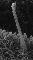
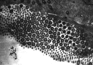
The cirrus is a bundle of individual cilia protruding from a single cell. This is illustrated when a cross section of the cirrus is viewed using TEM (below). These structures appear to be unique to sphaeriid and corbiculacean bivalves (Way, et al, 1989) and have not been observed in Unionacean bivalves thus far. The physical appearance of these structures also varies among species. For example, the cirri of Polymesoda caroliniana resemble horse tails, giving the appeance that the cilia of these structures are not bound together by any mechanism other than their common cell of origin. However, those of C. fluminea and some sphaeriid (fingernail) clams are much more organized in appearance as though the cilia that compose the currus were bound to one another.
Images of the cirrus and its cell are below.

1. An SEM of a cirrus on the gill of the Asiatic Clam (Corbicula fluminea, Muller 1774). Approximate magnification of the original photo – 3.3kx

2. TEM – Cross section through the cirrus near its base. Microvilli and ciliary structures are visible.

3. The cirri of Polymesoda caroliniana. The cirri are the long structures that resemble horse tails. Approximate magnification of the original photo – 1.03kx.
References Cited:
Way, CM, DJ Hornbach, T. Deneka, and RA Whitehead. 1989. A description of the ultrastructure of the gills of freshwater bivalves, including a new structure, the frontal cirrus. Can. J. Zool. 67:357-362.
