
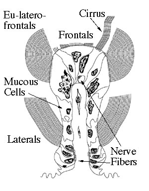
Below and to the left is a “clickable” idealized rendering of a gill filament of Corbicula fluminea,(Muller, 1774) the Asiatic Clam. Below and to the right is a transmission electron micrograph of the top portion of the gill filament. There are five major cell types that compose the ciliated epithelium of the gill of the Corbiculacean clams: the frontal cells, the cirrus cells, the eu-lateral frontal cells, the mucous cells, and the lateral cells. There are also nerve bundles within the filament of the gill. To see any of the structures mentioned above, you can click on the links above or you can click on any region of the idealized gill filament below and to the left. There are also micrographs of other structures of the gill available for viewing.
You can also click on any of the cell types (below in the image or above) to get a summary (and at least one electron micrograph) of its structure and function.
You can also click on any of the cell types (below in the image on the left or above) to get a summary (and at least one electron micrograph) of its structure and function.
|
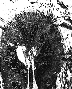 |
| TEM images were taken by Tony Deneka. SEM images were taken by Karen Moulton and Tony Deneka. | |