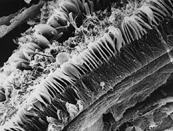
The lateral cilia are single cilia emerging from several cells near the base of the gill filament immediately beneath the mucous cell. These cilia are not bound together to form cirri as those of the eu-latero-frontal or the frontal cirrus cells. The cilia are thought to be the componant of the gill responsible for maintaining water flow through the gill for feeding and respiration (McMahon, 1991). These lateral cilia beat in metachronal waves. That is, the cilia beat in a wavelike and sequential manner starting from one end of the filament and proceeding to the other.
Below are images of he lateral cells and cilia.

1. An SEM of a lateral view of the gill filament of Musculium partemeium showing (from upper left to lower right) the frontal cilia, the eu-latero-frontal cirri, and the lateral cilia. Approximate magnification of the original photo – 1.08kx.

2. A TEM of the lateral cells of C. fluminea also showing the nerve fibers (small arrows) that may be involved in controlling metachronal wave activity. Also note the large number of closely packed mitichondria in this cell.
References Cited:
McMahon, RF. 1991. Mollusca:Bivalvia. Pp315-399. in</> Ecology and Classification of North American Freshwater Invertebrates. JH Thorp and AP Covich,eds.. Acedemic Press, Inc. New York.
