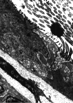
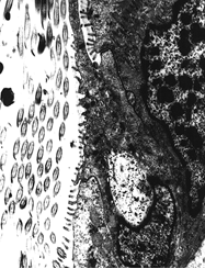
The mucous cell of the gill filament of Corbicula fluminea is situated between the eu-latero-frontal cell and the lateral cells. As the name suggest, this cell secretes mucous. Whether this mucous is an integeral part of the particle capture mechanism during feeding is not certain.
Below are images of mucous cells.

1. A TEM of a mucous cell of Corbicula fluminea. To the right are the cilia coming from lateral cells. You may be able to see the convoluted ER and secretory apparati that fill the interior of this cell.

2. A TEM of a mucous cell of Corbicula fluminea with a Eu-lateral-frontal cell toward the top and a lateral cell toward the bottom of the image. Visible is a mucous strand eminating from the cell. To the left are the lateral cilia in oblique section.
