
Eu-latero-frontal (ELF) cells are situated on the upper portion of the gill filament and form part of the arch of the filament. The ELF cirri, sometimes referred to as “plates,” are thought to be part of the filtering mechanism of the gill. The mechanism is composed of apposed ELF cirri extending toward one another to form a “net” of sorts (McMahon, 1991). Water is driven through this “net” by lateral cilia and the suspended particles are trapped and transferred to the frontal cilia where they are conducted to the food groove. There is some debate as to whether secreted mucous plays a significant role in the capture of particles.
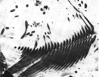
Below are some images of eu-latero-frontal cells and cirri.
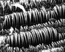
1. An SEM of the ELF cirri of Musculium partumeium. Also visible are one frontal cirrus (upper left quadrant). Approximate magnification of the original photo – 1.1kx.

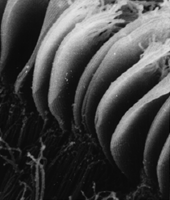
2. Detail of 1. The distal portions of many of the ELF cirri are visible, giving the cirri a “feathery” appearance. Approximate magnification of the original photo – 2.8kx.
3. A closeup of the ELF cirri of M. partumeium. Note that the dual sets of fused cilia that compose each “plate” are visible. Approximate magnification of the original photo – 5.2kx.
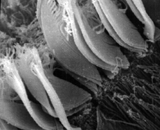
4. A closeup of the ELF cirri of M. partumeium. Again visible are the dual sets of fused cilia that compose each “plate.” Also visible in this micrograph are the distal, finger-like portions of the ELF cirri, the individual cilia that compose the cirrus, and the lateral cilia (bottom right quadrant). Approximate magnification of the original photo – 5.2kx.
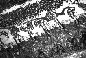
5. A TEM section through the gill filament of C. fluminea at the level of the ELF cells. This micrograph shows that the pairs of fused cirri that compose the “plates” issue from a single cell. A;so visible within this photo are the lateral cilia (a mass of cilia in cross section on the right side of the phto) and what appears to be a mucous strand (dark material running between the lateral cilia and the ELF cirri. Approximate magnification of the original photo – 3kx.

6. Detail of 5. This micrograph shows the pairs of cilia issuing from a single cell. In addition, this image shows that between each of the two “plates” of the ELF cirri, there is a single microvillus. Lastly, the image shows the ciliary rootlets that are bound to each cilium of the ELF. Also note that the rootlets on a given side of each of a pair of cilia cross to that side of the cell and end near the cells’ membrane.

7. A TEM of a section through the gill filament whose plane is at an approximate right angle to that shown in 5 and 6. In the lower half of the micrograph, at least one ciliary rootlet can be traced from the base of one of the cilia, across the cell, to the cell membrane. The roolets are visible as dark lines in this photo.
8. A TEM of a section in nearly the same plane as 6. This photo shows the fused cilia that compose the ELF cirrus.
References Cited:
McMahon, RF. 1991. Mollusca:Bivalvia. Pp315-399. in Ecology and Classification of North American Freshwater Invertebrates. JH Thorp and AP Covich,eds.. Acedemic Press, Inc. New York.