
The filament is the functional feeding unit of the Lamellibranch gill. It contains all of the cilliary structures required for respiration and food capture.
The images below are:
1. A cross section through the gill filament of Corbicula fluminea showing the frontal cells, the frontal cirrus cell, the eu-latero-frontal cells, and the mucous cell. Approximate magnification of the original photo – 3kx.
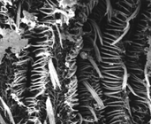
2. An SEM view of the gill of Corbicula fluminea showing the frontal cilia, the frontal cirri, and the eu-latero-frontal cirri. Approximate magnification of the original photo – 1.2kx.
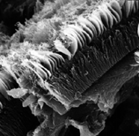
3. An oblique SEM view of the gill filament of Musculium partumeium showing the showing the frontal cilia, the eu-latero-frontal cirri, and the lateral cilia. Approximate magnification of the original photo – 1.04kx.
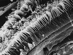
4. An SEM of a lateral view the gill filament of M. partumeium showing the frontal cilia, the plate-like eu-laterao-frontal cirri, and the lateral cilia. Approximate magnification of the original phot – 1.08kx.
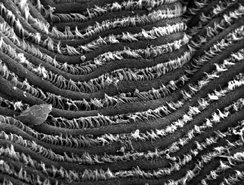
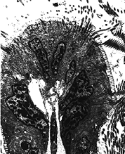
An SEM of the gill of Polymesoda caroliniana. Approximate magnification of the original photo – 1.03kx.