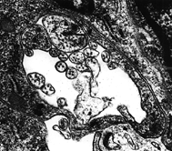
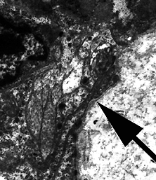
There are generally three separate bundles of nerve fibers that run through the gill filament of Corbicula fluminea. One set is generally assiciate intimately with the frontal cirrus cell. The other two are associated with the lateral cells. There is a larger bundle at the base of the lateral cell, and a smaller bundle nearer the eu-lateral-frontal cell. It is presumed that these nerve fibers associated with the lateral cells in some way arbitrate the metachronal wave activity manifested by the cilia of these cells. The function of the nerve fibers associated with the frontal cirrus cell, and why they are partitioned off from the other apical cells of the filament, is unknown at this time.
Below are images of the nerve bundles described above.

1. A TEM of the nerve fibers running along the base of the frontal cirrus cell of Corbicula fluminea. Note that the processes of the frontal cirrus cell completely surround the nerve fibers. These nerve fibers are isolated from the frontal cells and the eu-latero-frontal cells.

A TEM of the nerve fibers that are found associate with the lateral cells in the gill of Corbicula fluminea. This micrograph shows the nerve fiber bundle associated with the base of the lateral cell (arrow).
